
Presentation
A 70 year old female transferred from a private facility for investigation of abdominal pain 2 weeks post total knee replacement. On initial examination she was found to have a tense, painful abdomen with guarding. She was referred for surgical review and plain films were obtained.
Patient Data

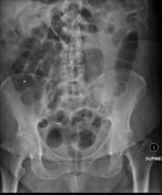
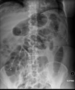
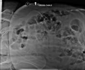
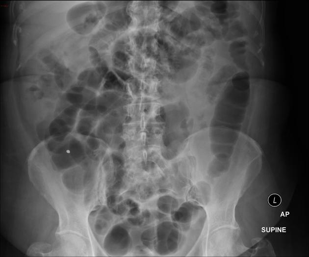
Plain films showed increased bowel gas but no evidence of obstruction, constipation, mass or perforation.


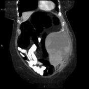

The surgical consultant examined the patient & was concerned about a palpable mass ordered a CT.
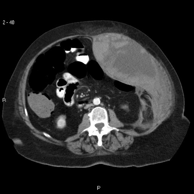
There is a large hematoma within the left anterior abdominal wall, involving the rectus abdominis, and the internal and external oblique muscles. This measures approximately 9cm x 20.9cm x 16.7cm. This is heterogeneous in attenuation likely due to presence of fibrin and blood products. There is contrast extravasation indicating active bleeding, within the superior abdominal wall (2-32 and 4-12), and within the mid abdominal wall posteriorly (2-46 and 4-22). Stranding is seen surrounding the hematoma.
The multilobulated cystic lesion involving the head and uncinate process of the pancreas appears unchanged in size since last scan, measuring approximately 51mm x 48mm x 52mm. The rest of the pancreas defines normally.


 Unable to process the form. Check for errors and try again.