
Presentation
Presents with 15 mm lower pole renal mass. Partial nephrectomy converted to total nephrectomy.
Patient Data
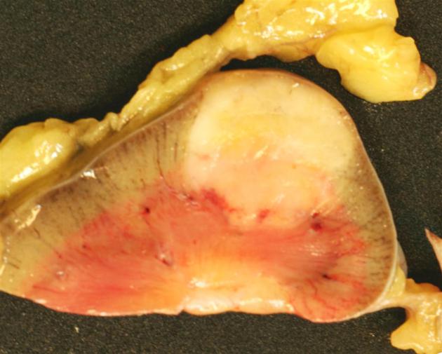
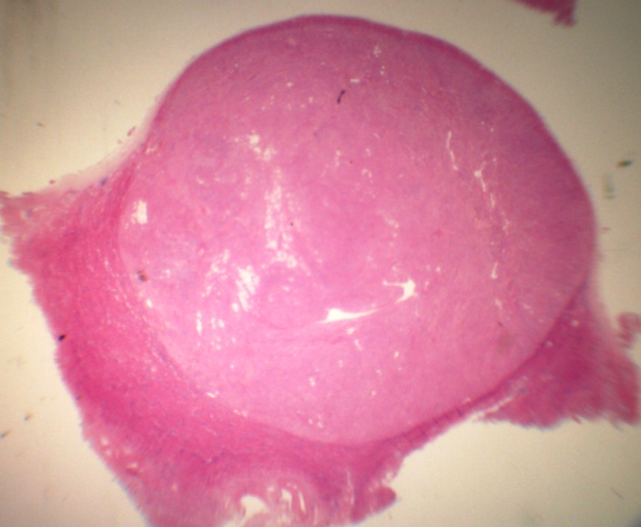
Macroscopic appearance of angiomyolipoma with predominantly pale grey to white tumor - there is a vague area of 'yellowish' tumor in the left upper quadrant corresponding to an area of rich in adipose tissue.

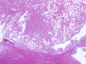
Low power histology of angiomyolipoma showing sharp circumscription. This is a muscle rich variant (solid eosinophilic areas dominate) with only scattered adipose tissue (interspersed cleared areas).
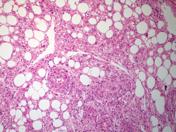
Medium power histology of angiomyolipoma with mixed smooth muscle and adipose tissue. There is a cuff of normal renal parenchyma in the lower aspect of the picture.
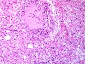
High power of angiomyolipoma with scattered thick walled vessels (central) set within stroma composed of smooth muscle (eosinophilic cells) and adipose tissue (clear rounded spaces).
Case Discussion
The textbook 'tri-phasic' appearance is not always present which can pose a diagnostic dilemma. As pathologists, we are seeing more core biopsies taken to try and identify the tumor type. On histology, the typical three components (vessels, smooth muscle and fat) are seen in variable proportions, but ‘monophasic’ (only one component) and ‘biphasic’ (two components) can be seen, especially if the tumor is only minimally sampled (e.g. core biopsy). Tumor cells are usually positive on immunohistochemistry for HMB45, CD117, CD63, and negative for cytokeratin and other epithelial markers.


 Unable to process the form. Check for errors and try again.