
Presentation
Three-day history of worsening right iliac fossa and flank pain with associated frank hematuria. Anemic with an acute kidney injury. Background of tuberous sclerosis with known renal angiomyolipomas.
Patient Data
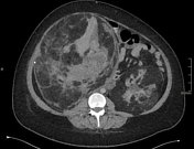


Large bilateral renal angiomyolipomas with minimal visible residual renal tissue. Hemorrhage within right renal angiomyolipoma with central hyperdense acute component and surrounding large sub-acute component extending inferiorly. Nil intraperitoneal hemorrhage/free fluid.
Distended gallbladder. Multiple hypodense liver lesions unchanged from previous imaging. Lung bases clear. No pleural effusion. Multiple sclerotic foci throughout the visible skeleton unchanged from previous imaging. VP shunt in situ.
Case Discussion
Renal angiomyolipomas are present within 55-75% of patients with tuberous sclerosis.The risk of spontaneous hemorrhage increases when tumor size increases beyond 3 cm in diameter.


 Unable to process the form. Check for errors and try again.