
Presentation
Flank pain and haematuria.
Patient Data
Age: 65 years
Gender: Male
From the case:
Renal calyceal diverticulum


Download
Info
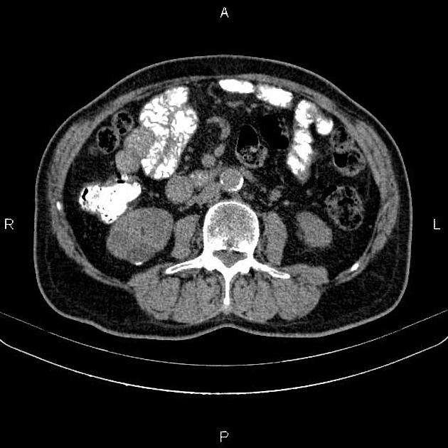
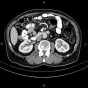
A 28mm cystic lesion is present at posterior aspect of right kidney, showing dependent dense calcific precipitate. This density increased in the delayed phase by excreted contrast accumulation inferring calyceal diverticulum. In addition, several non-enhanced simple cortical cysts are seen at both kidneys, with maximum diameters of 17mm. A few tiny stones, less than 2mm are seen at right kidney. A 4mm stone is also observed at lower calyx of left kidney.
Case Discussion
Calyceal diverticulum is a congenital outpouchings from the renal calyx or pelvis into the renal cortex and lined with transitional cell epithelium.


 Unable to process the form. Check for errors and try again.