
Renal calyceal rupture secondary to congenital pelviureteric junction obstruction
Presentation
Few days history of sudden onset epigastric and left sided abdominal pain
Patient Data


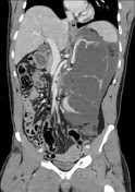
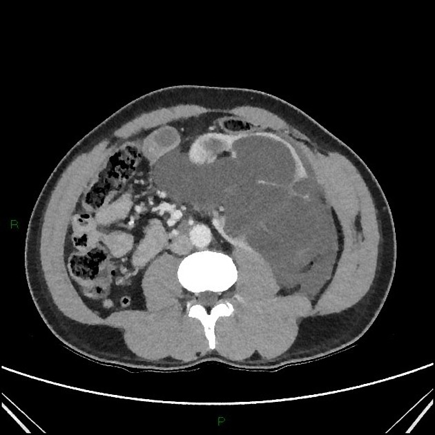
Severe hydronephrosis of the left kidney. Marked thinning of the renal cortex with evidence of rupture.
Perinephric collection with extension into the retroperitoneal space, tracking along the left paracolic gutter.
Beak-like narrowing of the dependant portion of the left renal pelvis without evidence of an obstructing calculus, lesion or distal hydroureter.
Case Discussion
This is a case of spontaneous renal rupture/pyelosinus backflow in an otherwise well individual with no previous medical history. There was no preceding trauma or presence of radiopaque renal calculi to suggest a cause of the injury. The severe global thinning of the left renal cortex is consistent with a chronic obstructive uropathy.
A stent was inserted under cystoscopy by urology. Overall imaging and intraoperative findings were consistent with congenital pelviureteric junction obstruction as a cause of the rupture.


 Unable to process the form. Check for errors and try again.