
Presentation
Prior breast cancer in 2015. Prior surgery for uterine leiomyosarcoma. Follow-up scan. Disease status?
Patient Data
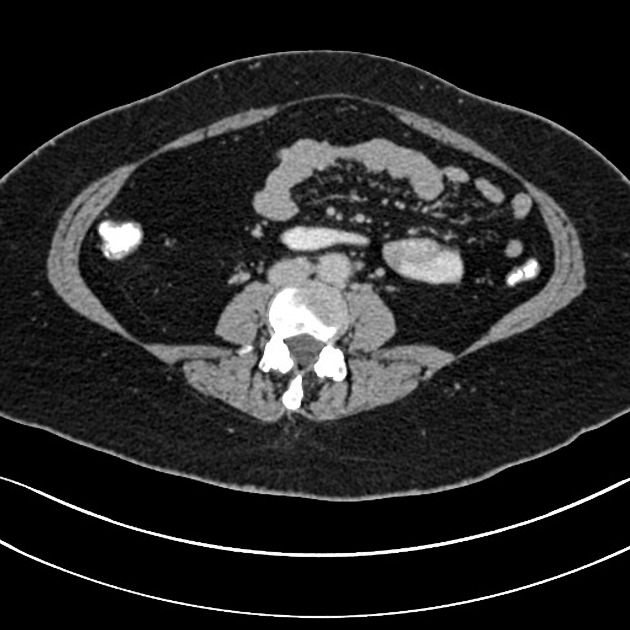
2016 scan. Performed for staging of breast cancer.
Hysterectomy and BSO.
2.1cm enhancing exophytic lesion arising from the lateral aspect of the midpole of the right kidney.
No para-aortic or renal hilar lymphadenopathy.
Fatty liver. Single small liver cyst.
Gallstones.
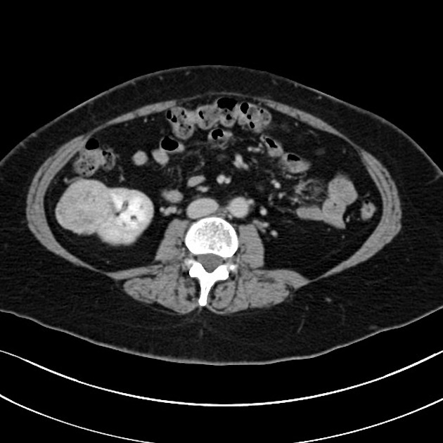
2018 FU scan. Renal lesion not mentioned in 2016 report.
4.4cm heterogenous mass arising from the lateral aspect of the midpole of the right kidney.
Fatty liver. Small liver cyst.
Gallstones.
No infradiaphragmatic lymphadenopathy.
Hysterectomy and BSO.
Case Discussion
With the overwhelming volume of CT imaging performed, incidental findings are very common. The majority are benign pathologies (e.g. gallstones, diverticulosis, renal cysts) but some are more sinister such as renal cell carcinomas and colonic malignancies. In fact, more than half of renal cell carcinomas are said to be incidentally found on imaging.
The differential in this context for the renal mass are:
- metastasis from breast carcinoma
- metastasis from leiomyosarcoma
- incidental renal cell carcinoma
It is hard to believe with a 2-year gap the former two are likely.
An ultrasound-guided biopsy was performed. The histology was one of renal cell carcinoma.
The identification of discrepancies should be shared and used for learning, hence the Royal College of Radiologists UK now calls these meetings.


 Unable to process the form. Check for errors and try again.