
Presentation
Renal mass on ultrasound.
Patient Data
Age: 55 years
Gender: Female
From the case:
Renal cyst - Bosniak I


Download
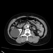
Info
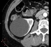
Hypoattenuating cysts characterized as homogeneous thin-walled and nonenhancing fluid-attenuation lesions.
From the case:
Renal cyst - Bosniak I
Download
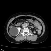
Info
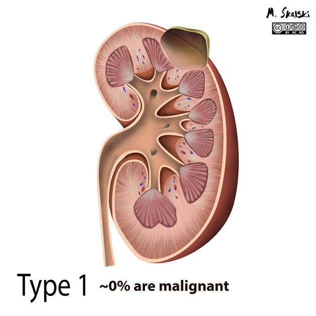
Illustration of the commonest type of Bosniak cyst - a type I ( simple cyst ).
Image credit: Matt Skalski
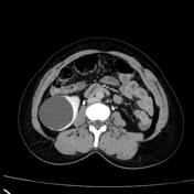
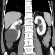
Case Discussion
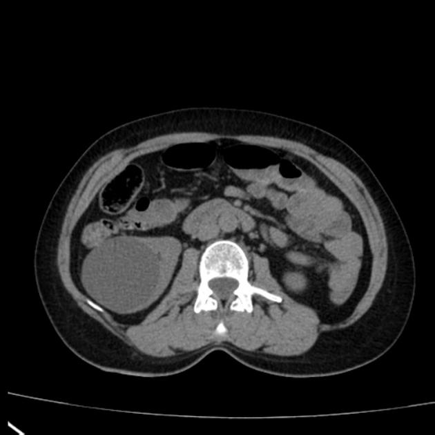
Hypoattenuating cysts, characterized as homogeneous thin-walled, non-enhancing fluid-attenuation lesions, are classified in Bosniak category I and are rarely problematic when encountered incidentally at contrast-enhanced CT.
Image Attribution
Author: Matt Skalski
Original file: https://radiopaedia.org/cases/bosniak-classification-of-renal-cysts-illustrations
License: Attribution-NonCommercial-NoDerivatives 4.0 International - Creative Commons
Modification: none


 Unable to process the form. Check for errors and try again.