
Presentation
Patient with long-standing uterine fibroids, increased ovarian tumor marker.
Patient Data
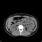

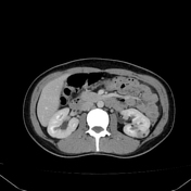

At the level of the interpolar region of the left kidney, there is a rounded exophytic lesion. The lesion is hyperdense in the non-contrast phase with some areas of fatty density, and enhances heterogeneously. More than 50% is exophytic, and it is close to the middle calyceal group. The collecting system is not dilated.
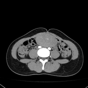
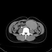
The uterus is heterogeneous with rounded hypodense lesions that show contrast enhancement similar to the uterus, suggestive of fibroids. Left adnexa with cystic components.
Case Discussion
Percutaneous fine-needle aspiration (PAAF) was performed, and anatomopathological studies showed predominantly haematous material with a group of connective tissue cells and tubules containing oncocytic cells.


 Unable to process the form. Check for errors and try again.