Presentation
Work up for haematuria.
Patient Data
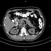

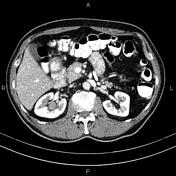
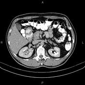

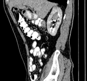

A 28 x 20 x 22 mm enhancing mass is present in the upper part of the right renal collecting system.
In addition, several non-enhanced simple cortical cysts are seen in both kidneys, with maximum diameters of 48 mm.
A small fat-containing umbilical hernia is present.
The prostate gland is enlarged.
Case Discussion
Pathology-proven right renal urothelial cell carcinoma without regional lymphadenopathy or detectable metastases.
Renal urothelial cell carcinoma is uncommon compared to renal cell carcinoma and can be challenging to identify on routine imaging when small. CT images show soft tissue density with only mild enhancement, usually significantly less enhancing than renal parenchyma or renal cell carcinomas.


 Unable to process the form. Check for errors and try again.