
Patient Data
Age: 75 years
Show annotations
Download
Info
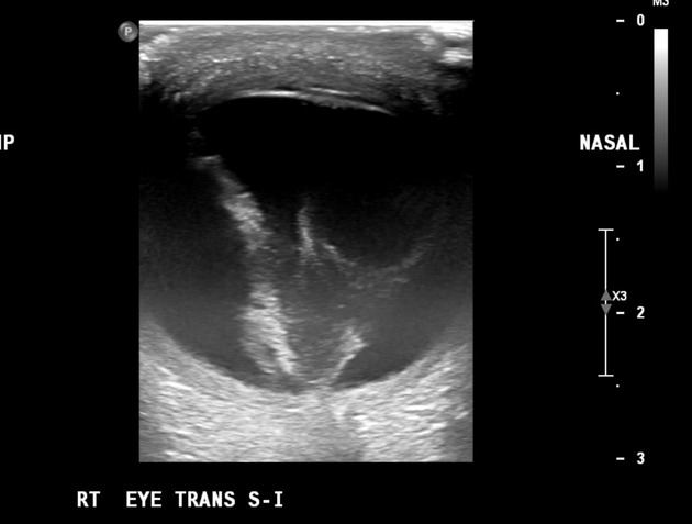
Membranes in the vitreous chamber with a somewhat triangular shape and insertion into the optic disc and ora serrata, typical for retinal detachment. There is also amorphous floating echogenic material in keeping with a component of hemo vitreous. The lens has increased echoes and slightly thickened walls in keeping with a cataract.
Show annotations
Download
Info
There is increased vitreous chamber density, with an identified triangular shape consistent with a retinal detachment.
Case Discussion
Typical imaging features of retinal detachment.


 Unable to process the form. Check for errors and try again.