
Presentation
Evaluate retroperitoneal mass.
Patient Data


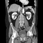
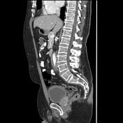
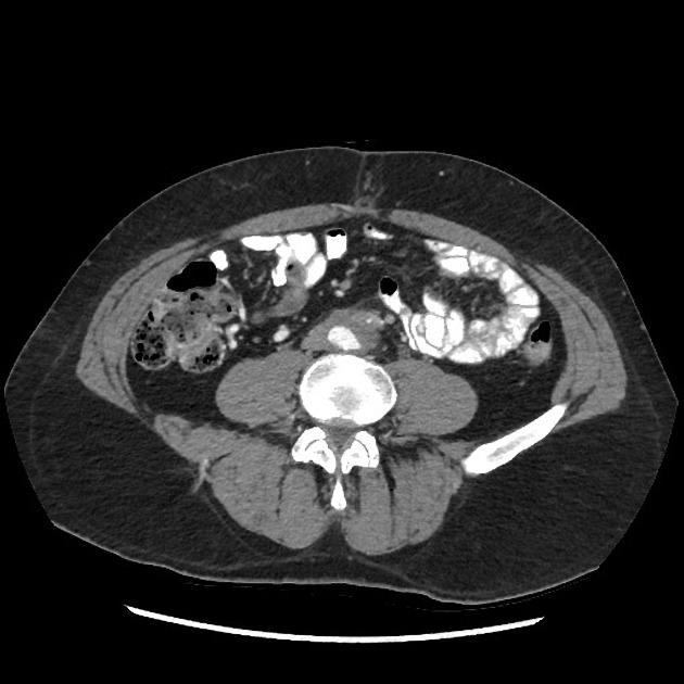
Circumferential soft tissue thickening involving the right internal iliac, bilateral common iliac arteries, and mid through distal abdominal aorta. Notice it surrounding the IMA. Both ureters are medialized but not obstructed. Aorta is not displaced anteriorly from the spine. No adenopathy. Transient small bowel intussusception left upper quadrant.
Case Discussion
Typical findings of retroperitoneal fibrosis with circumferential soft tissue surrounding the aortic bifurcation, iliac vasculature and distal descending aorta. It typically starts near the bifurcation and ascends. It is helpful to observe the medialization of the ureters which confirms that this is a fibrosing process. The lack of elevation of the aorta or adenoapthy makes lymphoma very unlikely. This could be confirmed with CT-guided biopsy or checking igG4 levels (which if elevated would obviate the need for biopsy, but if negative would not exclude RPF).


 Unable to process the form. Check for errors and try again.