Presentation
Old trauma, falling onto stretched hand 6 months ago.
Patient Data
Age: 65 years
Gender: Female
From the case:
Reverse Bankart lesion






Download
Info
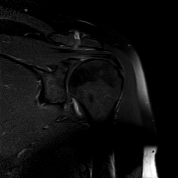
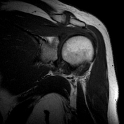
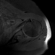
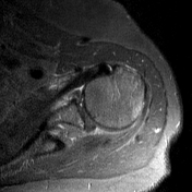
Well defined bony fragment at the posterior-inferior glenoidal surface, best seen on T1 fat saturated images
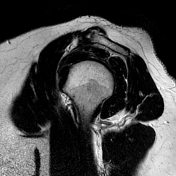
T2 and PDW increased and T1 decreased signals within AC joint reflects its inflammation
Heterogenous signals from thicker of supraspinatus muscle/tendon due to osteophytes from AC joint, leading to subacromial impingement, despite of acromion I type.
Subcoracoidal mild bursitis.
Case Discussion
This is uncommon case of the reverse Bankart lesion with subacromial impingement, arthritis in the AC joint.


 Unable to process the form. Check for errors and try again.