
Presentation
Rectal malignancy for staging
Patient Data


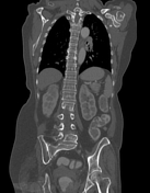
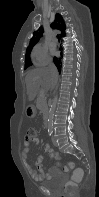
Diffuse osteopenia. Grade 1 anterolisthesis of L4-L5 and L5-S1. Lucent bands along the superior and inferior vertebral endplates with relative central sclerosis giving rise to the appearance of "reverse rugger jersey spine”
Eccentric lobulated lower rectal mass causing luminal narrowing. However, rectal contrast still able to pass through with no proximal bowel dilation. Several perirectal nodes.
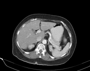
Other findings include fatty liver, gallstones, small right breast nodule, left subcutaneous abdominal wall lesion and multinodular goiter.
Case Discussion
Rugger jersey spine is usually seen in secondary hyperparathyroidism in which there are sclerotic bands involving the superior and inferior vertebral endplates with relative central lucency.
On the contrary, in reverse rugger jersey spine, there are instead lucent bands along the superior and inferior vertebral endplates with relative central lucency. In this case, these appearances may be due to underlying osteoporosis.


 Unable to process the form. Check for errors and try again.