
Presentation
Immunocompromised patient. Presenting with left facial pain and reduced sensation along V2 distribution. Rapidly developing left vision loss and complex ophthalmoplegia over a 24-hour period.
Patient Data
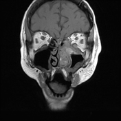


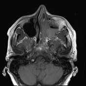
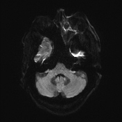


Note: MCA clip is likely the cause of the artifact degrading the quality of imaging in the cavernous sinus, orbital and left middle cranial fossa region.
Abnormal thickening/enlargment of left intraorbital segment of left optic nerve, left medial and inferior rectus muscles, which likely represents inflammatory changes. Restricted diffusion in the intraorbital segment of left optic nerve, suspicious of acute infarction.
Soft tissue is noted within the left orbital apex and anterior part of left cavernous sinus.
Significant fat streakiness within the left intraconal and extraconal fat, as well as the left retroantral fat.
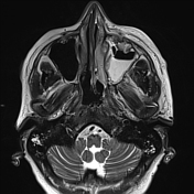
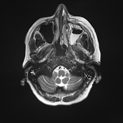
Almost complete fluid opacification within the left maxillary sinus and aerosolized secretion within the left sphenoid sinus.
Reduced mucusal signal intensities noted within the left nasal turbinates and left nasolacrimal duct (normally the inflamed mucosa would be hyperintensity on T2W sequence).
Irregular mucosal thickening within the left nasal cavity.
The MRI shows inflammatory changes that would be in keeping with an acute invasive fungal infection in context of the clinical history and imaging features.
The patient then underwent surgical debridement which showed necrotic tissue. Histopathology on tissues samples obtained showed fungal hyphae with microbiology confirming diagnosis of mucormycosis.
Case Discussion
Mucormycosis, or 'black fungus,' is an invasive infection seen in immunocompromised patients. The fungi are present in nature, originating from decaying vegetation and soil 1. Infection from mucormycosis is often rapidly progressive, involving angioinvasion leading to infarction and necrosis of host tissues.
A high mortality rate is associated with the infection, and even a diagnosis within 10 days of onset can carry a mortality rate of 80% 2.
In cases of rhino-orbital cerebral mucormycosis, MRI findings can include enlargement of the rectus muscles, inflammation of the optic nerve, as well as sinusitis 3-5, as was the case in this patient.
The most important imaging features are the presence of extra-sinus lesion or fat streakiness (such as in orbital apex, cavernous sinus) with concurrent sinusitis. If contrast is given, the presence of "black turbinate sign" is helpful for acute invasive fungal disease.
Early treatment with amphotericin B is indicated when infection is suspected, and surgical debridement is also often recommended 6.


 Unable to process the form. Check for errors and try again.