
Presentation
Sudden onset headache and meningism.
From the case:
Ruptured intracranial dermoid

Download
Info
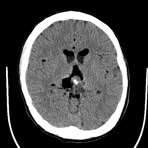
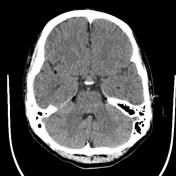
Two selected axial non-contrast CT images demonstrate hypodense material lying above the CSF in the frontal horns of the lateral ventricles, and around the pineal gland as well as smaller locules in the Sylvian fissures bilaterally and in the cerebellopontine cistern.
Case Discussion
The differential would be of air/gas or fat which can be distinguished on windowing and Houndsfield unit measurment. In this case, the material is confirmed to be of fat density, consistent with a ruptured intracranial dermoid.


 Unable to process the form. Check for errors and try again.