
Presentation
Acute lower abdominal pain, radiating to shoulder tips. Pregnancy test positive in ED but gestation unknown. US showed free fluid but otherwise inconclusive.
Patient Data
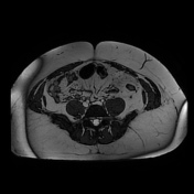

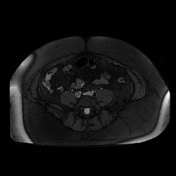

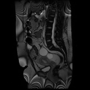
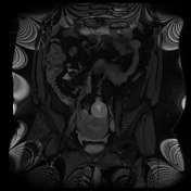

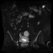
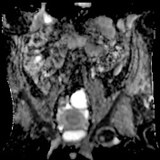

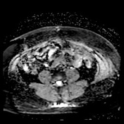
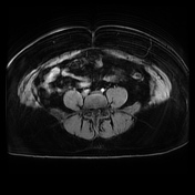
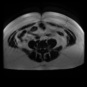
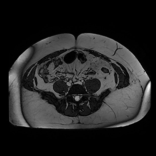
No intra-uterine gestation sac. Cysts arising from left ovary. Normal right ovary. Ovoid shaped structure anteroinferior to the right ovary with heterogeneous internal signal and a surrounding rim of intermediate signal, with this signal change extending into space anterior to the urinary bladder and posterolaterally towards the right iliac fossa. This intermediate signal exhibits restricted diffusion. Free fluid in posterior pelvis and in both paracolic gutters.
Findings in keeping with right-sided tubal ectopic pregnancy, with localized signal change indicating hematoma and therefore likely ruptured ectopic pregnancy.
Histopathology
Clinical History: Right salpingectomy for ruptured ectopic.
Macroscopic: A dilated Fallopian tube measuring 60 x 22mm. Separate hemorrhagic tissue measuring 40 x 30 x 15mm. No vesicles or foetal parts identified.
Microscopic: This Fallopian tube is distended and contains blood clot with occasional chorionic villi. These appearances are in keeping with the clinical impression of a tubal ectopic pregnancy. There is no evidence of gestational trophoblastic disease.
Conclusion: Right Fallopian tube - tubal ectopic pregnancy confirmed.
Case Discussion
Ultrasound is the modality of choice for the evaluation of abdominopelvic pain in pregnancy, with MRI being a useful alternative or second-line investigation when ultrasound is unavailable or due to inconclusive results. In this case, MRI has enabled confirmation of a tubal ectopic pregnancy, with no intra-uterine gestation sac, and a sentinel hematoma with widespread free fluid indicating rupture.


 Unable to process the form. Check for errors and try again.