
Scapholunate fracture with dorsal intercalated segmental instability
Presentation
Falling down with the outstretched left hand.
Patient Data


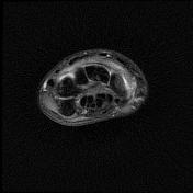
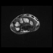
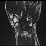
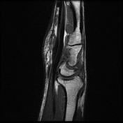
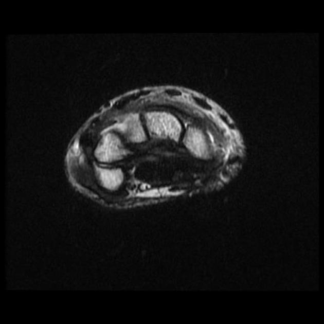
Small mid carpal joint effusion is seen.
Moderate dorsal intercarpal and radiocarpal ligament sprain.
Scaphoid waist fracture associated with marked bone marrow edema/bruise at the proximal and distal poles is present.
Congenital elongated deformity of the lunate bone associated with a fracture line at the lunate bone distal pole and adjacent bone marrow edematous changes are seen.
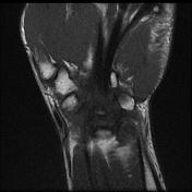
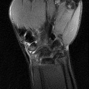
Dorsal intercalated segmental instability alignment as scapholunate angle 104° and capitolunate angle 32° is seen. Appearances may be exacerbated by the ulnar and slightly pronated wrist position.
Bone marrow edema/bruise also is seen at the other wrist proximal row bones and first metacarpal neck.
The fluid signal also is noted at the distal radioulnar joint.
Case Discussion
Scaphoid and lunate bones fractures account for ~65% (range 50-80%) and ~4% of all carpal bone fractures respectively. As mentioned, the lunate bone fracture is rare and in the literature, most cases were diagnosed almost before the era of CT and MRI.
Scaphoid fracture occurs essentially at any age, adolescents and young adults are most commonly affected. The scaphoid waist fracture is the most commonly affected site with possible avascular necrosis of the proximal pole. Carpal instability also can be seen as a complication of a scaphoid fracture.


 Unable to process the form. Check for errors and try again.