
Presentation
Cyclical lower abdominal pain with tenderness on palpation associated with the menstrual cycle. History of previous cesarian section two years ago
Patient Data


Ultrasound study shows a well-defined ovoid lesion of heterogeneous echogenicity measures 30 x 20 mm is noted closely related to the uterine previous cesarian section scar. It presents complex echogenicity with mixed hypoechoic echotexture and internal hyperechoic echoes. No detectable internal vascularity on the color duplex study.
Right ovarian small simple cyst measures 30x25 mm is also noted


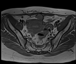
MRI study shows a well-defined ovoid lesion intraperitoneally at the midline of the pelvic region located between the rectus muscle and anterior inferior surface of the uterine body along the line of the previous cesarian section scar and closely related to the uterine scar. It measures 2.5x2.5x3 cm and elicits blood signal in the term of high signal in T1 & T1 fat sat and low signal in T2.
The lesion is seen closely related to the superior urinary bladder wall that appears irregularly thickened with internal minute foci of high signal in T2.
Thickening of the anterior uterine wall junctional zone measures 28mm and shows minute foci of bright T2 signal.
Left lateral uterine wall small myoma and right ovarian small simple cyst are also noted.
Case Discussion
Scar endometriosis can be located in the skin, subcutaneous tissue, rectus muscle/sheath, intraperitoneally, or in the uterine myometrium (within uterine scar). This case is an example of the intraperitoneal location.
In this case, the patient presented with a clinical history of cyclical lower abdominal pain with tenderness on palpation associated with the menstrual cycle that is almost typical clinical history for the endometriosis
Radiologically, the lesion is located along the surgical line of the cesarian section scar and is closely related to the uterine scar. It elicits a hemorrhagic signal that is one of the most common radiological presentations of endometriosis and is associated with uterine adenomyosis. All these features are highly suggestive of scar endometriosis
Ultrasound contribution by Dr/Samar El Sawy


 Unable to process the form. Check for errors and try again.