
Presentation
History withheld.
Patient Data


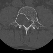

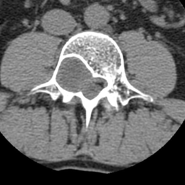
Large intra-extradural mass eroding the borders of the right neuroforamen and the dorsal contour of the L4 vertebral body. Marked rim sclerosis suggests a non-aggressive lesion.

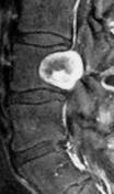
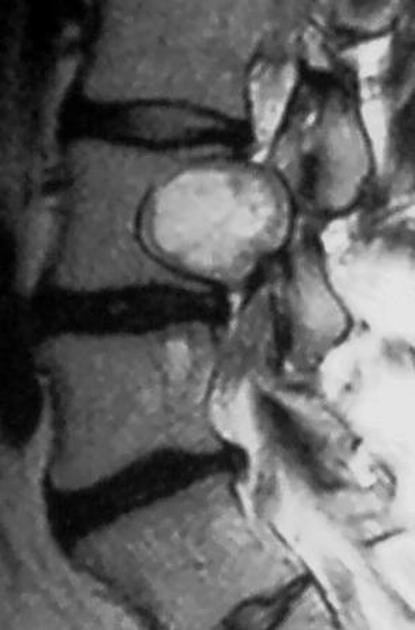
MRI shows a T2-hyperintense, somewhat heterogeneous lesion, that enhances avidly, but only at the periphery.
Case Discussion
Biopsy-proven Schwannoma at the L4 level on the right known for several years. The patient refused operative treatment.
In contrast with osteolysis, scalloping is characterized by a smooth margin with rim sclerosis towards the spinal canal. In the spine, it is caused by a mismatch between intra-spinal pressure and bone stability. Systemic disorders, that may lead to vertebral scalloping are connective-tissue diseases, mucopolysaccharidoses, neurofibromatosis type I or ankylosing spondylitis. Localized scalloping may also be caused by slowly growing intraspinal masses 1.
MR images courtesy of Dr. Christoph Gill.


 Unable to process the form. Check for errors and try again.