
Presentation
Chronic headaches. Blindness with enophthalmos on the right (previous trauma of right eye in childhood) and past history of surgery of the left eye 4 years ago.
Patient Data


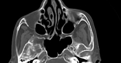
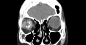
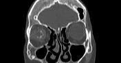
A high attenuation ring encircles the left globe in keeping with scleral buckle surgery.
Small retracted and atrophied right globe with dystrophic calcification in keeping with a phthisis bulbi.
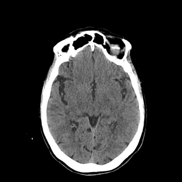
No parenchymal abnormality at the infra-or supratentorial level.
Normal size and configuration of the ventricular system.
Case Discussion
Scleral buckles are devices (often made from silicon) implanted into the eye for the treatment of retinal detachment. They are performed to relieve vitreous traction which closes the defects in the retina and reduces the recurrent detachment. They are usually permanent and removed only if there is a complication such as a device infection or extrusion.
Phthisis bulbi, also known as the end-stage eye, is an atrophic, scarred and disorganized non-functional globe which may result from severe ocular insults including injury.
Additional contributor/ Z.E Boudiaf, MD


 Unable to process the form. Check for errors and try again.