
Presentation
Known history of diffuse systemic sclerosis
Patient Data
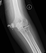

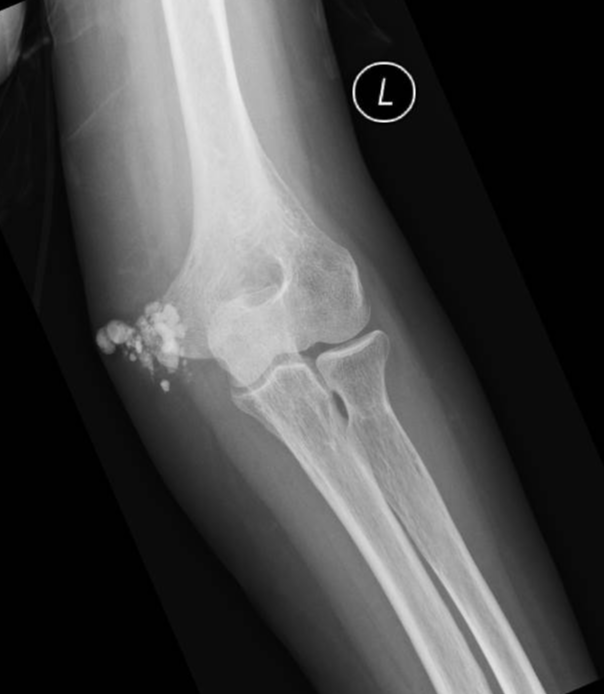
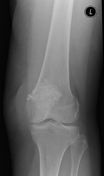
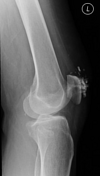
Periarticular soft tissue calcification seen posteromedially to the elbow joint.

Prepatellar soft tissue calcification.
Often seen over bony prominence, as in this instance.
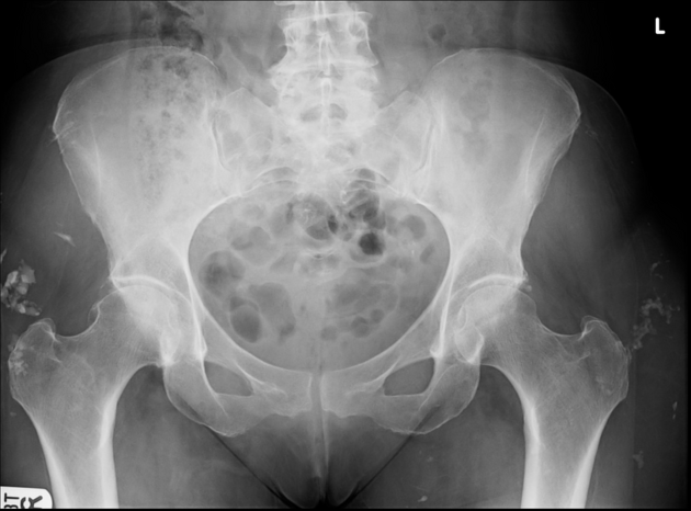
Amorphous calcification within the lateral soft tissues of the greater trochanters bilaterally.
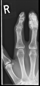
Partial resorption of the terminal tuft of the third distal phalanx (early acro-osteolysis).
Flexion contraction and soft tissue atrophy at the visualized interphalangeal joints.
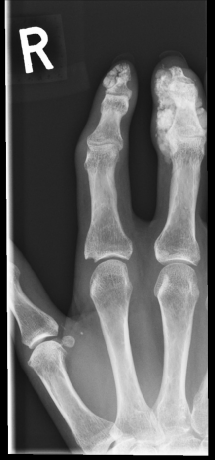
Extensive soft tissue calcification at the second and third phalanges.
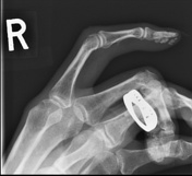
Erosion at the radial aspect of the base of the second proximal phalanx.
Case Discussion
Scleroderma is a multisystem autoimmune disease which can have many manifestations in multiple systems and organs.
Musculoskeletal changes can occur both in the soft tissue and bones. The hands are the most commonly affected site.
In this case, there are multiple sites affected principally demonstrating subcutaneous/periarticular calcification.


 Unable to process the form. Check for errors and try again.