
Presentation
Tip of thumb ulcer. ? osteomyelitis
Patient Data
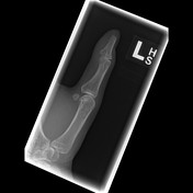
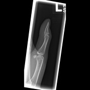
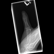

Erosion of the terminal tuft of the first distal phalanx. Given the clinical history of ulcer at this site, the appearance is suspicious for osteomyelitis.
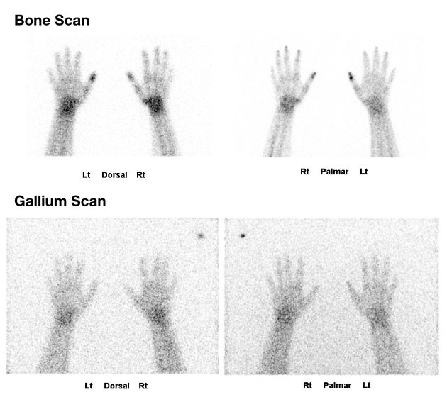
Increased uptake within the distal phalanx of the left and right thumb on the bone scan without any corresponding increased uptake on the gallium scan to suggest infection. The bilateral nature of the abnormality and lack of gallium uptake suggests a systemic cause for the active bone erosion.

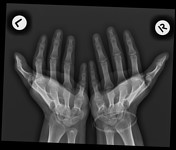
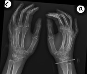
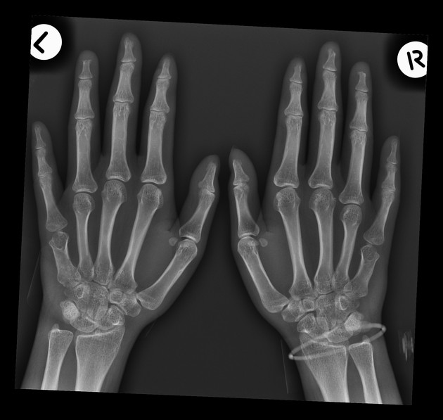
Bilateral first to fourth acro-osteolysis. No associated soft-tissue calcifications or evidence of erosive joint disease. Short fifth metacarpals bilaterally.
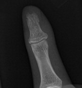
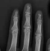

Zoomed images showing the acro-osteolysis.
Case Discussion
Acro-osteolysis due to scleroderma (confirmed subsequently). This case is interesting in that the presenting history and isolated radiographic appearance of the thumb was suspicious for osteomyelitis however the bilateral nature of the abnormality on bone scan and negative gallium scan ultimately led to the diagnosis of scleroderma with the identification of more extensive acro-osteolysis of other fingers. No cause for the short 5th metacarpals was identified.


 Unable to process the form. Check for errors and try again.