
Presentation
Right anterior neck swelling for 3 years, gradual increase in size with intermittent discharge from punctum.
Patient Data
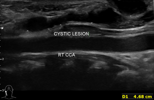
A tubular-shaped, cystic-like mass seen communicating from the right anterior neck skin extending superiorly anterior to the right common carotid artery. Unable to ascertain the extent of the superior course. No internal vascularity seen within. Multiple echogenic foci seen within this cystic mass which may represent infective component. No calcification or solid component seen within this mass.


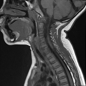
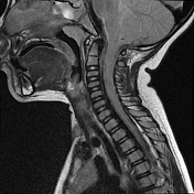


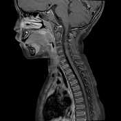
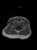
A well defined thin-walled tubular cystic mass at the right anterior neck where the inferior part of this mass has external punctum, found anterior and medial to right sternocleidomastoid muscle (T1 vertebral level).
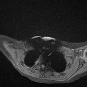
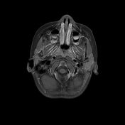
This tubular cystic mass travels deep to the right platysma muscle and it ends as blind ending sinus in between the right proximal internal and external carotid arteries (at C1 vertebral level). No internal fistulous communication with the right tonsillar fossa. [The course of the tubular mass is best assessed with thin slice T1 axial sequence, where it appears as dark signal intensity].
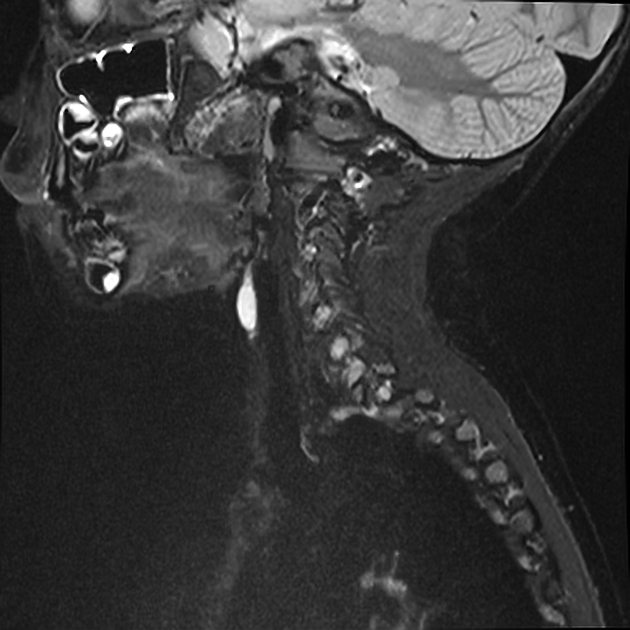
This cystic mass elicits hypointense on T1W and hyperintense on both T2W and STIR sequences. On post gadolinium administration, it demonstrates peripherally wall enhancement.
An enlarged cervical lymph node at right level II with multiple subcentimetre lymph nodes.
Case Discussion
The slightly greater degree of wall enhancement and echogenic foci within the tubular cystic mass in ultrasound, as well as right level II cervical lymphadenopathy suggest the ongoing infection of second branchial cleft sinus.
Second branchial cleft anomaly (cyst, fistula and sinus) accounts for 95% of all branchial cleft anomalies.
Cysts can occur anywhere along the course of the second branchial apparatus, from the pharyngeal wall to the skin. The external punctum is found anterior and medial to the sternocleidomastoid (SCM) on the neck skin.
It passes laterally and inferiorly between the internal and external carotid arteries. The angle of the mandible is a common location.
Bilateral second branchial cleft cysts can be associated with branchio-oto-renal syndrome.
According to Bailey classification (1929), this case is to be classified as type III: extends between the internal and external carotid arteries.


 Unable to process the form. Check for errors and try again.