
Presentation
Severe abdominal pain and distension with inability to pass stools for 2 days and urine for 1 day.
Patient Data
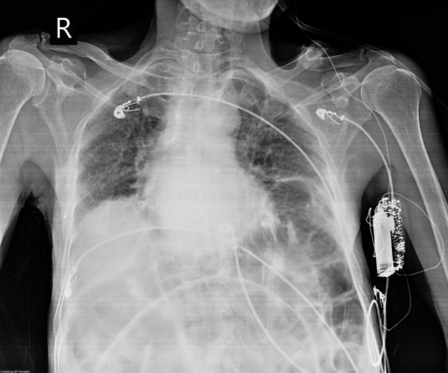
Supine radiograph shows the apex of a grossly dilated large bowel loop.


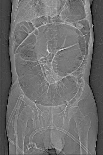
Topogram shows a grossly dilated large bowel with lack of haustrations. Coffee bean sign is seen. The dilated colon points towards the pelvis.
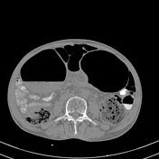
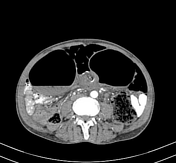
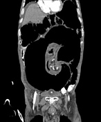
Grossly dilated sigmoid colon with multiple air-fluid levels forming a closed loop associated with swirling of mesentery at transition point seen.
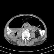
Tapering obstruction at the rectosigmoid junction obstructing proximal passage of rectal contrast medium.
Other findings: Mild ascites, small left inguinal hernia and right inguinal hernia containing bladder bilaterally and loculated fluid on the left. Bilateral renal cortical cysts, and prostatomegaly.

Intra-operative image shows grossly dilated sigmoid colon with a twist point and vascular congestion.
Case Discussion
This is a classical case of sigmoid volvulus. The patient presented with severe abdominal pain and distension with inability to pass stools for 2 days and urine for 1 day.
He had consumed antispasmodics with no relief. Patient was admitted and a supine AP view radiograph was taken which revealed dilated bowel loops.
Contrast enhanced CT scan of abdomen was performed which showed the classical radiological signs of sigmoid volvulus.
Within a few hours, the patient was taken up for surgery. Intra-operative image is attached above. Surgery was successful and the patient recovered well.


 Unable to process the form. Check for errors and try again.