
Presentation
Bilateral lacrimal gland masses
Patient Data


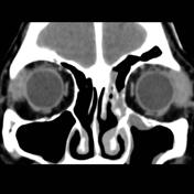
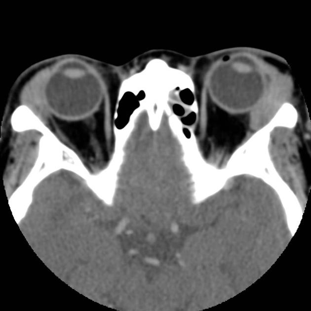
Bilateral lacrimal gland masses with homogeneous enhancement.
Case Discussion
Sjögren syndrome: presenting with bilateral lacrimal gland masses
Final diagnosis
Biopsy, left lacrimal gland. Reactive lymphoid hyperplasia, consistent with Sjögren syndrome.
Microscopic description
The left lacrimal gland biopsy shows a heavy lymphoid infiltrate with destruction of the acini and preservation of ducts. The infiltrate is composed of large lymphoid follicles with prominent and irregularly shaped germinal centers surrounded by a small lymphocytic infiltrate. Immunoperoxidase stains show prominent B cell staining of the follicles with T cells in the interfollicular space. Staining for light chains shows equal numbers of kappa and lambda light chains. The histologic changes are most consistent with the inflammatory infiltrate seen in Sjögren syndrome.


 Unable to process the form. Check for errors and try again.