Presentation
Headache
Patient Data


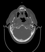
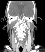
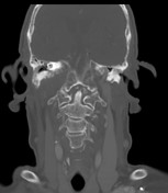
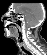

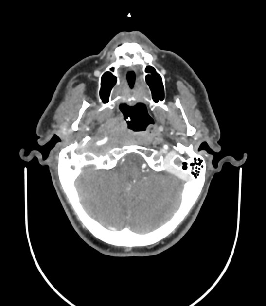
There is an abnormal soft tissue thickening involving the right carotid, prevertebral and parapharyngeal spaces. This is associated with erosions and osteolysis of the right base of the skull including the clivus, petrous apex and anterior wall of the carotid canal. This abnormal soft tissue causes compression of the proximal internal jugular vein and narrowing of the petrous portion of the right internal carotid artery. There is also mass effect on the right aspect of the nasopharynx, however, the nasopharyngeal mucosal space appears normal with no evidence of abnormal masses.
There is opacification of the right mastoid air cells and middle ear cavity with mild soft tissue thickening of the external auditory canal.
No neck lymphadenopathy detected.
Case Discussion
CT findings are consistent with right skull base osteomyelitis (SBO) secondary to right otomastoiditis.
DD:
- skull base metastasis
- nasopharyngeal carcinoma
Types of SBO:
- typical: secondary to necrotizing external otitis (NEO), petrous apicitis or otomastoiditis
- atypical (Central): usually secondary to invasive sinusitis, deep infection or could be idiopathic


 Unable to process the form. Check for errors and try again.