
Presentation
Slowly growing frontal swelling of the skull for some time.
Patient Data
Age: 20 years
Gender: Female
Note: This case has been tagged as "legacy" as it no longer meets image preparation and/or other case publication guidelines.
From the case:
Skull vault hemangioma


Download
Info

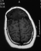
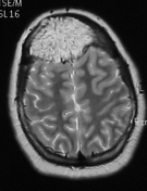
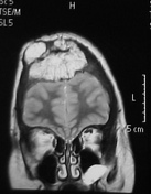
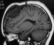
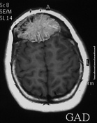
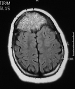
An expanding lesion is seen in the frontal bone of the skull vault. It appears low signal on T1 and high signal on T2 & FLAIR sequences. It shows intense homogenous enhancement in the post-contrast study. It has the appearance of sun-ray trabecular pattern.
Case Discussion
The MRI findings are compatible with skull vault hemangioma.


 Unable to process the form. Check for errors and try again.