
Presentation
Nodular left frontal region swelling for many years
Patient Data
Age: Young
Gender: Male
From the case:
Soft tissue hemangioma

Download
Info
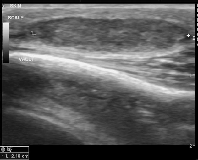
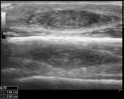
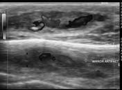
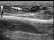
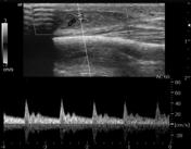
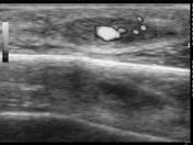
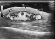
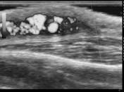
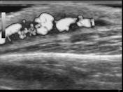
A well-defined, solid, ovoid lesion which measures 21 x 18 x 5 mm is seen in the subcutaneous plane. No bone erosion is noted. No calcification or cystic changes. Significant flow is noted on color Doppler with an artery entering and exiting the lesion.
Case Discussion
Surgical excision - histopathology: hemangioma.


 Unable to process the form. Check for errors and try again.