
Presentation
Headache.
Patient Data


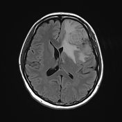
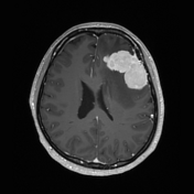
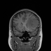
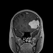
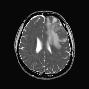
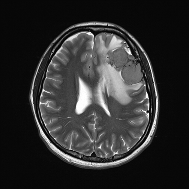
A large multilobulated extra-axial mass in the left frontal region is isointense on T1 and T2 weighted images to the adjacent cortex and has vivid contrast enhancement.
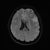
The posterior component demonstrates diffusion restriction with ADC values similar to brain (~750 x 10-6 mm2/s) whilst the anterior component of the mass predominantly demonstrates facilitated diffusion, although there are tiny patchy areas of diffusion restriction at its centre. This implies differing cellularity.
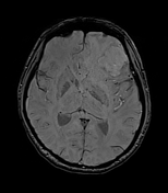
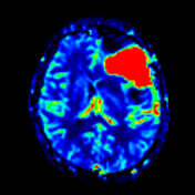
Prominent vascularity is seen within the lesion. MR perfusion demonstrates significantly increased cerebral blood volume.
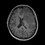
Surrounding hyperintense FLAIR signal in keeping with cerebral oedema. Mass effect with effacement of the left lateral ventricle and rightward midline shift of 5mm. Mild/early left uncal herniation. No tonsillar herniation. No hydrocephalus. No other abnormal lesions.
Conclusion
Large enhancing vascular extra-axial left frontal tumour, likely a meningioma, although there are multiple atypical features e.g. marked lobulation and vascularity over the surface of the mass. Variant/higher grade meningioma or solitary fibrous tumour are possible differentials.
Case Discussion
The patient went on to have a biopsy.
Histology
Sections show a highly cellular tumour attached to dura. The tumour is composed of a relatively uniform proliferation of ovoid spindle cells in haphazard fascicles, with intervening thin walled staghorn-shaped vessels. There are focal areas of lower cellularity, with interspersed dense keloidal-like collagen. Mitoses are relatively brisk, up to 6 per 10 HPF (2.5/mm2). There is no necrosis.
Immunohistochemistry
STAT6: Moderate to strong patchy nuclear positivity
CD34: Diffuse membranous positivity
EMA: Negative SSTR2: Negative
GFAP; Negative
Ki-67: Up to 20% in hotspots.
Final diagnosis: solitary fibrous tumour, CNS WHO grade 2.


 Unable to process the form. Check for errors and try again.