
Presentation
Progressive paraplegia
Patient Data
Age: 35 years
Gender: Female
From the case:
Spinal arteriovenous malformation


Download
Info
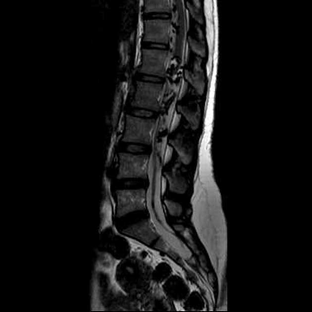
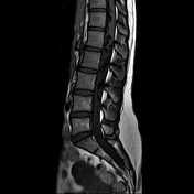
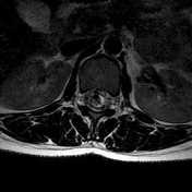
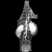
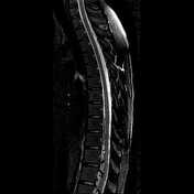
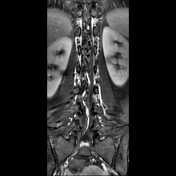
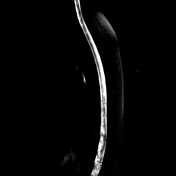
MRI demonstrating conus medullaris edema and enlarged perimedullary and infraconal vessels. MRA MIP depicted the feeding artery of the lesion to derive from the eighth thorasic level (T8) on the left side. Localization of the shunt is at dural level.
Case Discussion
Lumbar spine MRI shows appearance of a spinal dural arteriovenous malformation. Progressive myelopathy from venous congestion/hypertension of dural AVM due to delayed diagnosis, leading to Foix-Alajouanine syndrome.


 Unable to process the form. Check for errors and try again.