
Patient Data


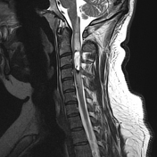
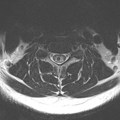
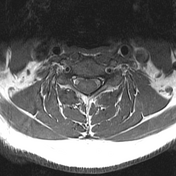
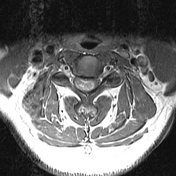
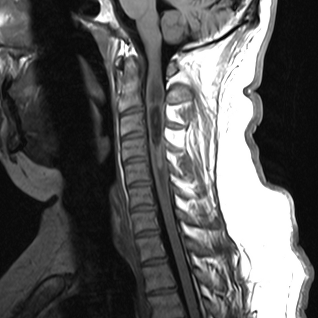
Extending from C2/3 to the midpoint of C4 is an intramedullary mass which is of high T2 signal, low T1 signal and demonstrate prominent contrast enhancement (seen on axial imaging). It has a prominent hemosiderin capping with some intrinsic high T1 signal consistent with hemorrhage. A moderate amount of vasogenic edema extends above and below the lesion from the crater cervical junction to C7. The remainder of the cervical cord, the cervical vertebrae, and paraspinal soft tissues are unremarkable. The vertebral arteries are codominant to demonstrate normal flow voids.
The patient went on to have a laminectomy and excision of the mass.
Histology
Sections show a moderately cellular glial tumor. The tumor comprises a mixed population of cells, some showing glial features with fibrillary processes, yet others displaying more epithelioid features. Perivascular pseudorosettes are a prominent feature. No mitotic figures are identified and there is no necrosis or endothelial cell hyperplasia. The tumor cells are strongly GFAP positive, show punctate dot like cytoplasmic staining for EMA, and membranous staining for CD99. The Ki-67 proliferative index is approximately 1%.
Final diagnosis
The morphological and immunohistochemical features are of a spinal ependymoma (WHO Grade II)
Case Discussion
This case illustrates typical and characteristic appearances of a spinal ependymoma with prominent hemosiderin capping.


 Unable to process the form. Check for errors and try again.