
Presentation
2 weeks of postural headache.
Patient Data


Of note, these images were obtained 30 minutes after a contrast-enhanced MRI brain.
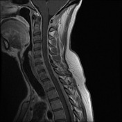
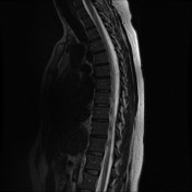
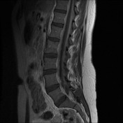
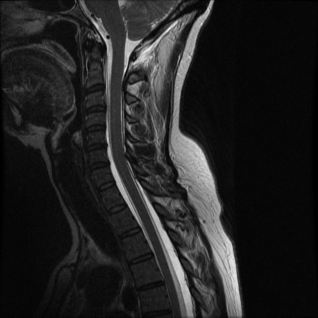
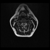
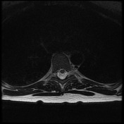
An extensive extradural fluid collection involving the entire spinal canal partially effaces the thecal sac. Posteriorly at C1-C2 fluid is also seen in the soft tissues (C1-C2 false localizing sign).
The collection is T2 and T1 hyperintense to CSF and contains multiple engorged epidural veins. The signal hyperintensity may be due to proteinaceous content or due to contrast accumulation.
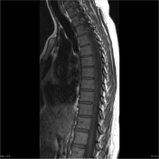
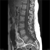
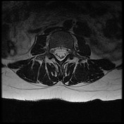

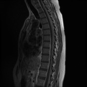
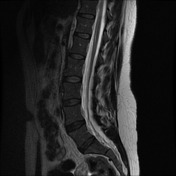
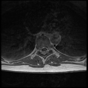
MRI spine was performed the following day, to obtain non-contrast enhanced T1 sequences and to complete the study with T2 axial sequences.
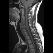
In the absence of contrast, the extensive epidural collection is confirmed to be isointense to CSF on T1 and T2 sequences.

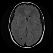
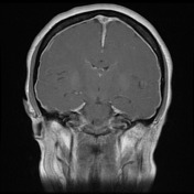
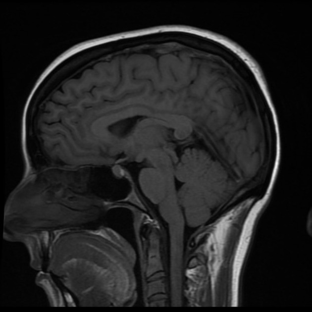
Diffuse smooth enhancing pachymeningeal thickening, enlargement of dural venous sinuses and plump pituitary gland are all typical features of intracranial hypotension. Non-specific T2 hyperintense focus in left frontal white matter is incidental to the presenting complaint.
Case Discussion
The spinal manifestations of spontaneous intracranial hypotension are infrequently described and most often encountered when a spinal MRI is performed to search for a site of CSF leak.
Typically, MRI reveals an extensive nonfocal extradural CSF collection (spinal hygroma) that is isointense to CSF and contains dilated epidural veins. The dural sac usually maintains its midline attachment to the posterior longitudinal ligament with the collapse of the anterolateral thecal sac, giving a 'festooned' appearance. Smooth circumferential dural enhancement is common. Occasionally, delayed contrast leak into the collections has been described (as in this case).
Importantly, it is common to see a 'collection' of CSF between the spinous processes of C1 and C2. This is the C1-C2 false localizing sign and should not be mistaken for the site of the CSF leak.


 Unable to process the form. Check for errors and try again.