
Presentation
Four weeks of bilateral posterior pleuritic chest pain.
Patient Data

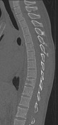
Lungs and pleural spaces are clear. Cardiomediastinal contour is normal. Anterior wedging of T10 vertebral body with irregularity of the inferior endplate and left paraspinal opacity. Absent right L1 pedicle (winking owl sign).


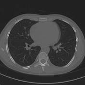
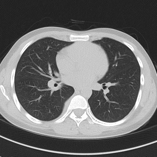
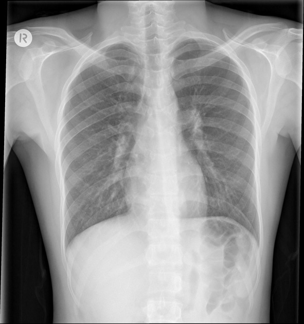
Lungs and pleural spaces are clear. No mediastinal lymphadenopathy. Upper abdomen is unremarkable.
T10 crush fracture with vertebral body lytic lesion. Destructive lytic lesion at the right L1 vertebral body involving the right pedicle.
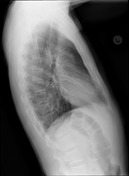
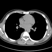
Right seventh rib lytic lesion with adjacent soft tissue density.
The patient underwent image guided aspiration of the paraspinal soft tissue:
DETECTED NUCLEIC ACID TESTING (ON SPECIMEN) DNA Amplification Assay for M.tuberculosis Complex (PCR): See comment below M. tuberculosis Complex PCR: DETECTED
POSITIVE MYCOBACTERIAL CULTURE 1. Acid Fast Bacilli ISOLATED
Case Discussion
Spinal tuberculosis (Pott disease) is not an uncommon manifestation. In this case, the presentation and imaging are non-specific, and the imaging differential diagnosis includes other lytic bone lesions such as metastases and lymphoma.


 Unable to process the form. Check for errors and try again.