
Presentation
Left hypochondrium pain and fever.
Patient Data
Age: 60 years
Gender: Male


Show annotations
Download
Info
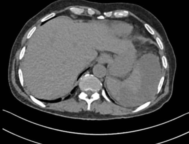
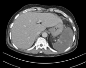
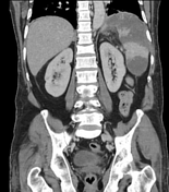
Moderate splenomegaly with multiple confluent non-enhancing hypodense areas in the splenic parenchyma, reaching up to the periphery. Mild surrounding perisplenic fat stranding/ inflammatory changes were seen. The splenic vein and splenic artery appear normal.
Case Discussion
The patient's WBC count was 24,910/microlitre with a neutrophil of 95%.
The clinical history, imaging findings and lab parameters are suggestive of splenic infarction with superimposed infection.
The patient improved after supportive treatment & IV antibiotics and discharged in the stable condition.


 Unable to process the form. Check for errors and try again.