
Presentation
A male presented with acute abdominal pain. He has a history of coronary artery bypass grafting (CABG) surgery.
Patient Data


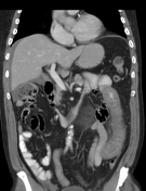
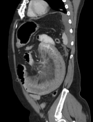
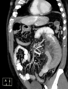
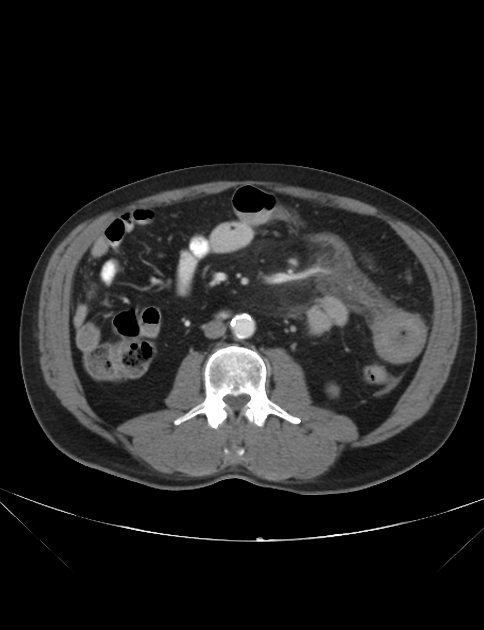
CT reveals abnormal diffuse segmental wall thickening at the level of the jejunum (long segment, 18-20 cm) on the left side. There is mesenteric oedema and vascular congestion, and some ascites.
Superior mesenteric artery (SMA) and vein (SMV) are patent, and no thrombus was seen.
Findings are most suggestive of non-occlusive mesenteric ischaemia and submucosal haemorrhage. Differential diagnosis includes Crohn disease and infectious enteritis.
Case Discussion
The patient underwent surgery. Surgery confirms submucosal jejunal haematoma on left with haemoperitoneum.
The patient has a history of coronary artery bypass grafting (CABG) surgery and was taking anticoagulation medication. The absence of vascular occlusion and filling defect on CT imaging makes differentiation of ischaemia from intramural haematoma or inflammation difficult.
A combination of history, physical examination, and imaging findings are all important in establishing the correct diagnosis.


 Unable to process the form. Check for errors and try again.