Patient Data
Age: 65 years
Gender: Male
From the case:
Spontaneous osteonecrosis of the knee
![](https://prod-images-static.radiopaedia.org/images/31955/84b2cf63d7520a568444f7285d1abc_thumb.jpg)
![](https://prod-images-static.radiopaedia.org/images/31956/a5cd7454b8bd86a25dec996891e36f_thumb.jpg)
Download
Info
![](https://prod-images-static.radiopaedia.org/images/31955/84b2cf63d7520a568444f7285d1abc_big_gallery.jpg)
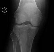
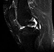
Oval radiolucency with central sclerosis in the subchondral medial femoral condyle.
From the case:
Spontaneous osteonecrosis of the knee
![](https://prod-images-static.radiopaedia.org/images/31902/0eab6481ffe7f66eee04be9695f3f6_thumb.jpg)
![This study is a stack](/packs/stack-YQKLCKBI.gif)
![](https://prod-images-static.radiopaedia.org/images/31913/fb0199938a81fef45c73ca39ab0ca5_thumb.jpg)
![This study is a stack](/packs/stack-YQKLCKBI.gif)
![](https://prod-images-static.radiopaedia.org/images/31921/09c1d2707f97f072cd7bbeab9b4575_thumb.jpg)
![This study is a stack](/packs/stack-YQKLCKBI.gif)
![](https://prod-images-static.radiopaedia.org/images/31945/caad96789037dee5f4ea7b02ed1643_thumb.jpg)
![This study is a stack](/packs/stack-YQKLCKBI.gif)
Download
Info
![](https://prod-images-static.radiopaedia.org/images/31902/0eab6481ffe7f66eee04be9695f3f6_big_gallery.jpg)
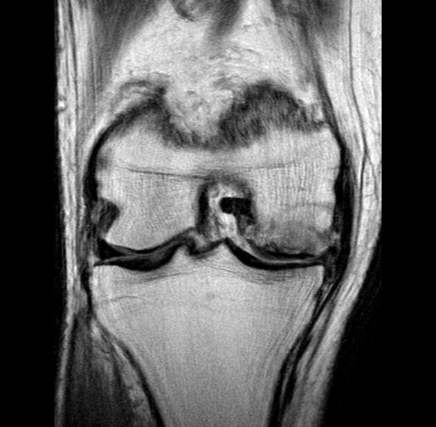
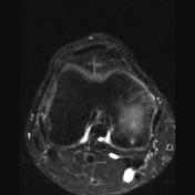
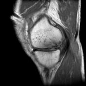
Spontaneous osteonecrosis of the knee (SONK) involving the weight-bearing portion of the medial femoral condyle, with associated articular surface impaction fracture. Maceration and degenerative tears of the body and posterior horn of the medial meniscus is associated with meniscal extrusion and osteophytic lipping. Incidental Bakers cyst.
Case Discussion
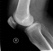
Spontaneous osteonecrosis of the knee (SONK) of the lateral aspect of the medial condyle (most common location).