Presentation
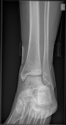
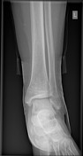
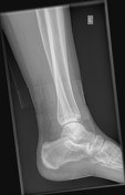
Ankle pain. No trauma. Normal x-ray.
Patient Data
Age: 35 years
Gender: Female
From the case:
Stress fracture - distal fibular
Download
Info

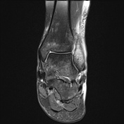
No fracture. No focal osseous lesion. No periosteal reaction. Alignment is normal.
From the case:
Stress fracture - distal fibular

Download
Info

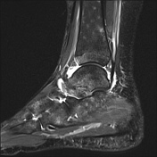
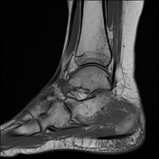
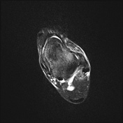
Florid marrow edema of the distal fibular shaft extending into the medial malleolus and marked adjacent soft tissue hyperintensity. Linear hypointensity through the medial distal fibular just above the ankle joint that incompletely extends laterally. Small ankle joint effusion. Patchy bone marrow edema may reflect disuse or CRPS.
Case Discussion
Typical MRI appearances of a stress fracture, although in an uncommon location - the inciting events for this patient are not known. In retrospect, and with the eye of faith, a sclerotic line in the same position as the fracture on the MRI can be seen.


 Unable to process the form. Check for errors and try again.