
Presentation
Amputation of the left upper limb for rhabdomyosarcoma of the forearm 4 years ago. Presented with stump pain and numbness.
Patient Data
Age: 13 years
Gender: Male
From the case:
Stump neuromas - upper limb

Download
Info

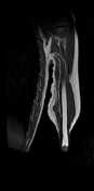
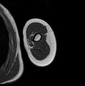
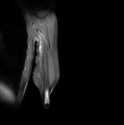
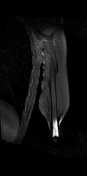
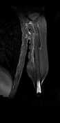
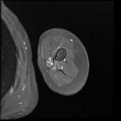
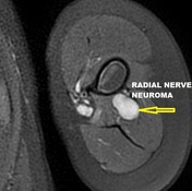
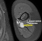
There are three well-defined fusiform masses along the course of the radial nerve (12 x 10 x 8 mm), median nerve (16 x 11 x 9 mm) and ulnar nerve (16 x 11 x 7 mm). They elicit an isosignal to the muscles on T1, high signal on T2 and STIR with moderate peripheral enhancement.
From the case:
Stump neuromas - upper limb
Download
Info
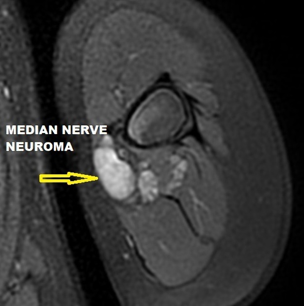
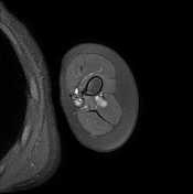
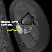
Annotated images of the median, radial, and ulnar nerves neuroma.
Case Discussion
MRI features are most consistent with stump neuromas arising from the end of the radial, median, and ulnar nerves.


 Unable to process the form. Check for errors and try again.