
Presentation
Present with anterior neck pain and odynophagia.
Patient Data
Age: 50
Gender: Male
Download
Info
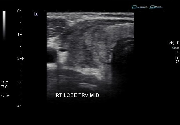
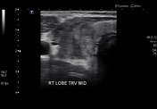
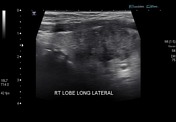
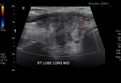
Ultrasound of the right lobe of the thyroid demonstrates an ill-defined irregular region of heterogeneous hypoechogenicity without elevation of flow on color Doppler examination.
Images courtesy of Dr Anthony Upton.

Download
Info
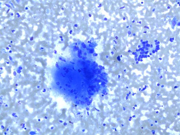
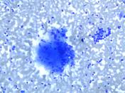
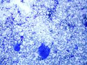
The FNA smears show an inflammatory process with features consistent with subacute (de Quervain's) thyroiditis:
- scattered benign reactive follicular epithelial cells
- prominent inflammatory background including multinucleate giant cells
- clusters of epithelioid histiocytes, consistent with granulomatous inflammation
Multinucleate giant cells and granulomas can be difficult to differentiate morphologically (see third image) but should not be confused theoretically:
- multinucleate giant cells are single cells with multiple nuclei, formed by the fusion of numerous macrophages (histiocytes)
- granulomas are aggregates of numerous 'epitheloid histiocytes' (activated macrophages).
Case Discussion
This case illustrates imaging and cytological features consistent with subacute granulomatous thyroiditis.


 Unable to process the form. Check for errors and try again.