Presentation
The decedent is a 30 year old male that had sustained a gunshot wound (GSW) to the head that impacted the skull base, fracturing it. The patient did not survive and was sent for autopsy.
Patient Data

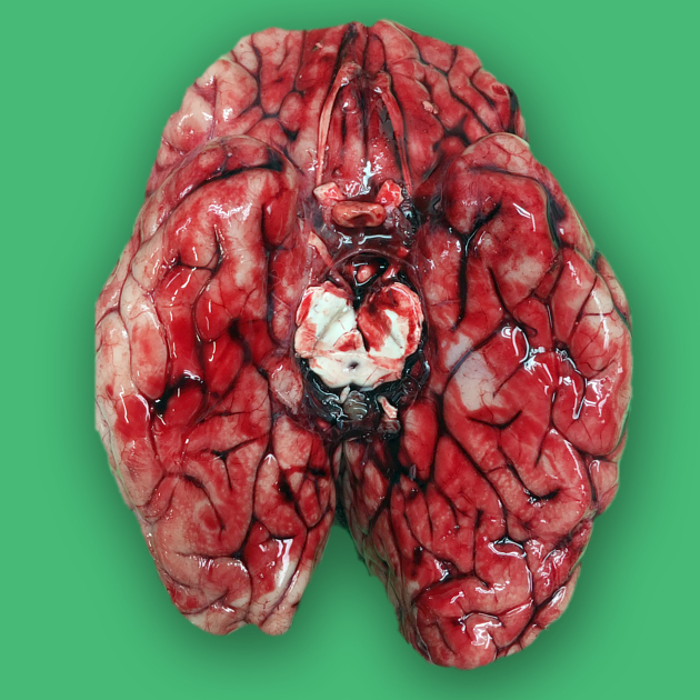
At the time of autopsy, the brain has been removed, the brain stem cut at the level of the upper cervical spinal cord. To best demonstrate the Circle of Willis, the cerebellum is the then removed with a cut through the midbrain and turned over to show the inferior surface. Extensive subarachnoid blood is visible filling the sulci. While some fresh blood is always present upon removal of a brain, the blood cannot be washed off the brain as it is located deep to the arachnoid layer which is very thin and transparent. This subarachnoid hemorrhage did not have a localized source and likely was due to shockwave impact of the bullet through the skull base.


 Unable to process the form. Check for errors and try again.