
Presentation
Right forefoot pain.
Patient Data

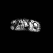
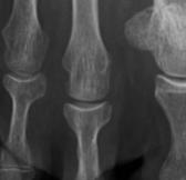
Moderate joint effusion, hyperemia, and synovial thickening in the right 2nd metatarsophalangeal joint. There is flattening of the dorsal articular surface of the 2nd metatarsal head, compared to the contralateral 2nd metatarsal head (4th image).
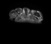

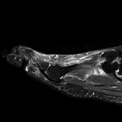
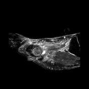
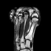
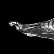

Confirmation of the US findings. In addition, there is bone marrow edema in the metatarsal head of the right 2nd metatarsal bone, with proximal extension to the metaphysis and the diaphysis. Dorsal subchondral bone hypointensity and cortical flattening. Mild-moderate periarticular soft-tissue edema.

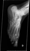
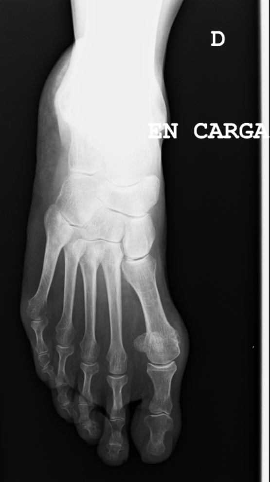
There is mild flattening and bone sclerosis in the lateral aspect of the 2nd metatarsal head.
Case Discussion
In this case, the diagnosis of subchondral insufficiency fracture was first diagnosed on ultrasound. Subchondral insufficiency fractures are frequently diagnosed on plain radiographs, MRI, or bone scans. Nevertheless, this diagnosis should be considered on ultrasound. Bone shape, synovitis, and soft-tissue inflammatory changes can be assessed by ultrasound with precision.
This is a case of subchondral insufficiency fracture, probably complicated with osteonecrosis. It is important to keep in mind that, despite this being the typical location of Freiberg disease, this entity implies osteochondrosis evolving into osteonecrosis. Therefore, it is only seen in children and adolescents who are growing. This is a middle-aged woman, so the term Freiberg disease cannot be used.


 Unable to process the form. Check for errors and try again.