
Presentation
Seizures.
Patient Data
Age: 17 years
Gender: Female


Download
Info
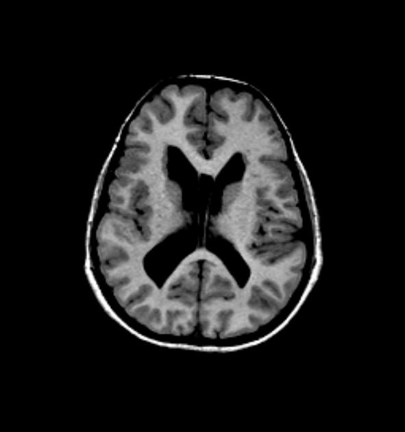
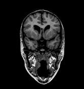
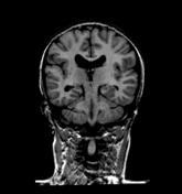
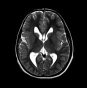
MRI of the brain demonstrates periventricular (subependymal) grey matter heterotopia best seen bulging into the frontal horn of the right lateral ventricle. Additionally the perisylvian grey matter is thicker than normal with nodular grey-white matter junction. Incidental note is made of a cavum septum pellucidum et vergae.
Case Discussion
This case demonstrates both subependymal grey matter heterotopia and polymicrogyria (a recognized association), in the context of a young woman with Turner's syndrome.


 Unable to process the form. Check for errors and try again.