
Presentation
Headaches
Patient Data


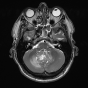
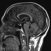
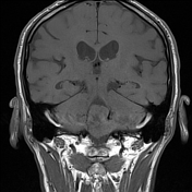
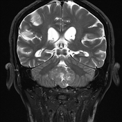
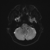

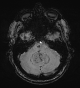
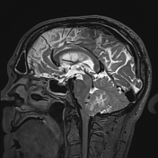
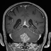
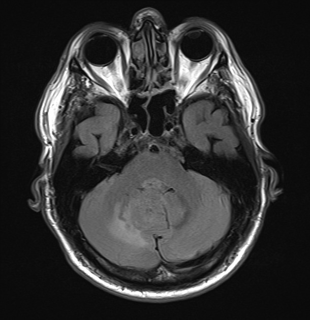
A large heterogeneous mass in the posterior fossa centred inferiorly either pushes the vermis superiorly (most likely) or arises from it. it is separate from the brainstem. The mass enhances vividly, and has facilitated diffusion compared to the adjacent brain, with multiple foci of blood product within it (note the larger ones have aliasing artefact).
Despite its size, and mass effect on the fourth ventricle, the third and lateral ventricles appear normal without obstructive hydrocephalus.
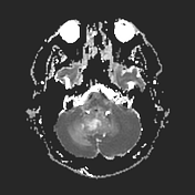
Within the pons is a capillary telangiectasis, faintly enhancing with profound SWI signal loss.
Conclusion:
In this age group and in this location and unusually large subependymoma is a distinct possibility. An meningioma arising form the edge fo the foramen magmum or falx cerebelli is a possibility. In the absence of known systemic malignancy, metastastic disease is less likely. Ependymoma, medulloblastoma, choroid plexus papilloma are all unlikely given demographics and imaging appearances.
Case Discussion
The patient went on to have a resection.
Histology
Sections show a tumour comprising cells with clustered small ovoid nuclei and prominent fibrillary background. There are also regions of calcification, microcysts and some hyalinised blood vessels. Mitoses, necrosis and nuclear atypia are not seen. There is no microvascular proliferation.
By immunohistochemistry, tumour cells are positive for GFAP and 02-40, while negative for EMA, olig2, SSTR 2 and sox 10. H3K27me3 shows intact nuclear staining and Ki-67 proliferative index is less than 5%.
Final diagnosis: subependymoma, CNS WHO grade 1.


 Unable to process the form. Check for errors and try again.