
Presentation
Mass in right submandibular gland.
Patient Data
Age: 45 years
Gender: Male
From the case:
Submandibular pleomorphic adenoma


Download
Info

Ultrasound demonstrates a well circumscribed hypoechoic mass in the right submandibular gland, measuring 1.0 x 1.2 x 1.4cm in diameter. Color examination demonstrates prominent internal vascularity.
Images courtesy of Dr Anthony Upton


Download
Info
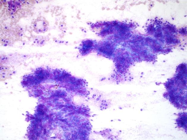
The FNA smears show classic features of pleomorphic adenoma:
- cellular, with single to irregular groups and sheets of epithelial cells
- interspersed stromal fragments
- background population of myoepithelial cells - often described as having a 'plasmacytoid' appearance
FNA submandibular gland (pap stain) - The smears show large numbers of epithelial cells interspersed with vaguely fibrillary to whispy stroma.
Case Discussion


 Unable to process the form. Check for errors and try again.