
Presentation
Pelvic pain, dysmenorrhea
Patient Data


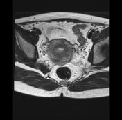
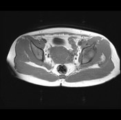
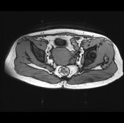


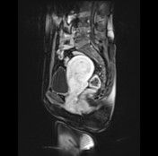
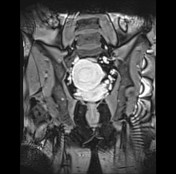
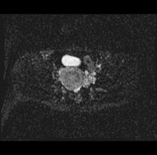
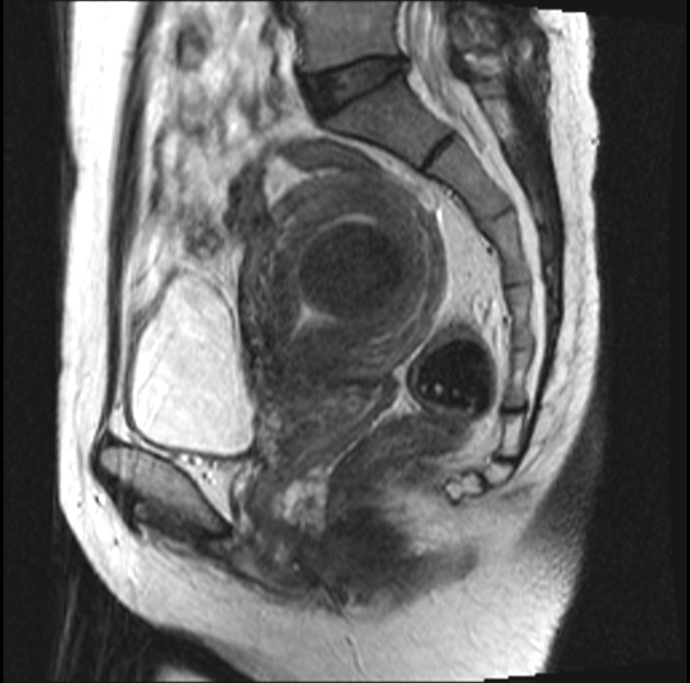
A submucosal mass is seen in the posterior wall of the uterus and displacing the endometrium anteriorly. It shows the homogeneous slight hypointense signal compared to myometrium on T2, the isointense signal on T1, enhancement on T1 C+, no restricted diffusion on DWI.
The hysterectomy was performed, histopathology confirm leiomyoma of the uterus.
Case Discussion
This case illustrates the submucosal uterine leiomyoma typically.
An imaging differential diagnosis includes endometrial polyps, which arise within the endometrium and can cause enlargement of the endometrial cavity, showing homogeneous echogenic and single vascular pedicle on ultrasound, isointense signal compared to endometrium on T1. As opposed to polyps, submucosal fibroids can displace or indent the endometrium, elicit hypoechoic masses with shadowing and multiple feeding vessels on ultrasound, low signal intensity on MRI.


 Unable to process the form. Check for errors and try again.