
Presentation
Hip pain.
Patient Data
Age: 16 years
Gender: Female
From the case:
Supra-acetabular fossa


Download
Info
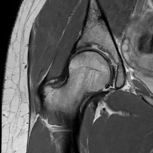
Focal 3mm deep 8mm wide fossa within the superior aspect of the acetabulum with cartilage continuing to line the fossa consistent with a supra-acetabular fossa (a normal variant). No evidence of any adjacent marrow reaction which you would certainly expect if this region represented an acquired osteochondral defect.
From the case:
Supra-acetabular fossa
Download
Info
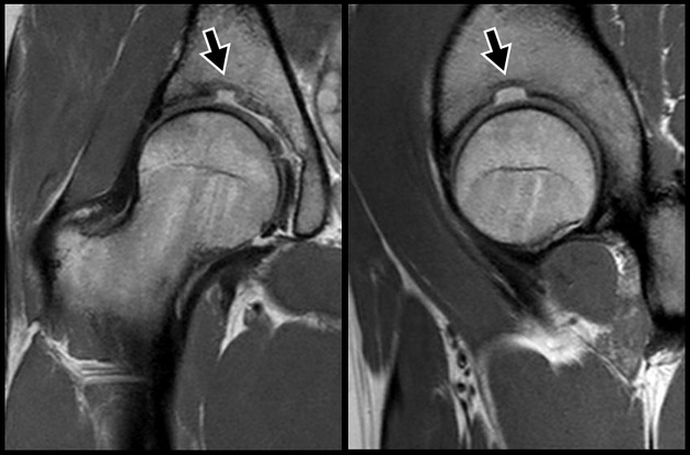
Coronal and sagittal PD weighted images of a supra-acetabular fossa. Note how the fossa remains lined with intermediate signal cartilage.
Case Discussion
Characteristic MRI appearance of a supra-acetabular fossa which is a normal variant. There is a small hip joint effusion which may indicate synovitis as a cause for the patient's symptoms.


 Unable to process the form. Check for errors and try again.