
Presentation
Post-operative examination immediately following excision of posterior fossa mass
Patient Data
Age: 65 years
Gender: Male
From the case:
Susceptibility artifact on brain MRI due to gas bubbles

Download
Info
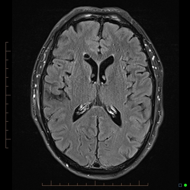
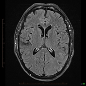
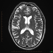
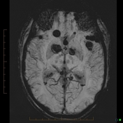
Multiple low signal intensity rounded filling defects with in the subarachnoid space and lateral ventricles anteriorly. These are poorly seen on the FLAIR sequence, better seen on T2-weighting, and "bloom" on the SWI sequence.
Case Discussion
It is important to recognize artifacts on MRI in order to avoid error in reporting. The clinical history in this case is important as well as the distribution of the signal alteration (floating anteriorly with the patient in a supine position).


 Unable to process the form. Check for errors and try again.