Presentation
Knee pain and discomfort. No history of trauma. No other joint affection.
Patient Data


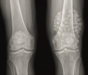
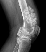
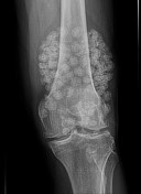
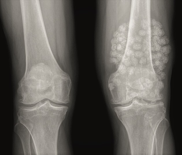
Multiple well-defined, rounded, "lamellated" opacities of almost similar sizes noted confined to the left knee joint space. No evidence of fascial or muscular extension. No bony erosions. Concentric calcifications in the form of ring, arcs and swirls noted.
Mild degenerative changes in the medial compartment with early osteophyte formation.
Case Discussion
The descriptive pattern of calcifications confirms the chondroid origin of this disease. Findings are of primary synovial osteochondromatosis of the knee joint. This appearance is so specific with the commonest location being in the knee; that confident diagnosis can be made without the need for biopsy.


 Unable to process the form. Check for errors and try again.