Presentation
History of treated syphilis was admitted to the emergency department with fever and bacteremia. Analysis of cerebrospinal fluid shows an increased protein level and lymphocytic pleocytosis.
Patient Data
![](https://prod-images-static.radiopaedia.org/images/52951599/9012814bdcd89a9214944b7520046e7d5d8f7cb1580a38db167d46264e9b00df_thumb.jpeg)
![This study is a stack](/packs/stack-YQKLCKBI.gif)
![](https://prod-images-static.radiopaedia.org/images/52951612/079230c09cd7b7b23d5babdc482fc6ea52c24e218997e731ba65ddb411aebee7_thumb.jpeg)
![This study is a stack](/packs/stack-YQKLCKBI.gif)
![](https://prod-images-static.radiopaedia.org/images/52951626/e7c1e4485426a0ebb17a62d2651ac900a60ab841bcd480b613b2705017994ba7_thumb.jpeg)
![This study is a stack](/packs/stack-YQKLCKBI.gif)
![](https://prod-images-static.radiopaedia.org/images/52951638/2fc4c8860246b5c0d4976fc1ed6a65aebf547fcfe8e3b6153bf1ccf2085ae275_thumb.jpeg)
![This study is a stack](/packs/stack-YQKLCKBI.gif)
![](https://prod-images-static.radiopaedia.org/images/52951657/5e7113a77defd4dca53580bfbfb1209d574c0981ebbf23d597656fb30cb3aed0_thumb.jpeg)
![This study is a stack](/packs/stack-YQKLCKBI.gif)
![](https://prod-images-static.radiopaedia.org/images/52951687/59a175ce1a3a470c024b9c80193a480f5456741dfc1a74f5708e9b0e8e65e18e_thumb.jpeg)
![This study is a stack](/packs/stack-YQKLCKBI.gif)
![](https://prod-images-static.radiopaedia.org/images/52951599/9012814bdcd89a9214944b7520046e7d5d8f7cb1580a38db167d46264e9b00df_big_gallery.jpeg)
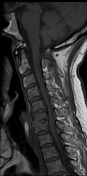
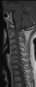
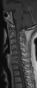
Saggital T2 and STIR images show high signal that affects the dorsal columns of the cervical spinal cord at the C5-C6 level. Neither enhancement of the lesion on post-contrast sequence nor hemorrhagic focus is shown.
A slight leptomeningeal enhancement is associated throughout both anterior and posterior medullary surfaces.
![](https://prod-images-static.radiopaedia.org/images/52951669/3d8c5fca90976d372cc672f6334cc38aae2be11f30fabcb375d8840c157cc7fd_big_gallery.jpeg)
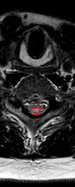
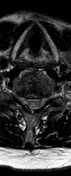
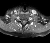
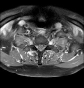
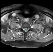
Axial T2 image shows bilateral and symmetrical hyperintensity within the dorsal columns of the spinal cord (red circle).
![](https://prod-images-static.radiopaedia.org/images/52951700/3732ab45856ee9170acd59938474c5e18d05c0c904c2a8e98aeb800f9b896c3d_thumb.jpeg)
![](https://prod-images-static.radiopaedia.org/images/52951701/3c2bca711ae1fac5b1b2b6a306bc1872371870957b55648c6c5772fdfa7bf8ab_thumb.jpeg)
![](https://prod-images-static.radiopaedia.org/images/52951702/45e5307ff40a1a456b59c66ad98ec41954ce261a0b4b97865b488cce66a74d74_thumb.jpeg)
![](https://prod-images-static.radiopaedia.org/images/52951703/19c96d73ced7664397eae3df1e82c8ed42543bb83a99810ae13d1a043a01cd98_thumb.jpeg)
![](https://prod-images-static.radiopaedia.org/images/52951700/3732ab45856ee9170acd59938474c5e18d05c0c904c2a8e98aeb800f9b896c3d_big_gallery.jpeg)
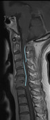
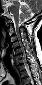
Saggital and axial T1 post-contrast images show a slight leptomeningeal enhancement in the anterior and posterior medullary surfaces.
Case Discussion
Tabes dorsalis appears as a late manifestation of late tertiary neurosyphilis, resulting in demyelination of posterior columns of the spinal cord. Most usually, there is symmetrical and bilateral hyperintensity on T2/STIR images. Generally, these areas do not enhance.
The principal differential imaging diagnosis to consider includes:
- multiple sclerosis: generally asymmetrical with a slight extent
- transverse myelitis: usually not confined to dorsal columns
- HIV-related vacuolar myelopathy: history of HIV infection needed
- neoplasm: not particularly of dorsal columns; cord expansion; often enhance
- ischemia: acute onset, restriction on DWI
The presence of specific antibodies to Treponema and the absence of other microorganisms in the analysis of cerebrospinal fluid guides the possibility of acute reinfection in this case.
This case is submitted in collaboration with Drs. Ana M Quiles Granado and Nerses Nersesyan.