
Presentation
Known patient (Asian) of Takayasu arteritis presented to the ER with acute onset right sided weakness
Patient Data
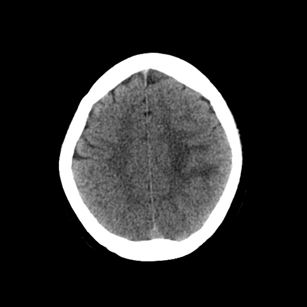
Effaced cortical sulci involving left superior parietal lobule with subcortical and deep white matter hypoattenuation confined to the pre and post central gyri. No focal hemorrhage or midline shift.
Rest of the neuroparenchyma is unremarkable.
An MRI was recommended to assess in detail.
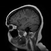

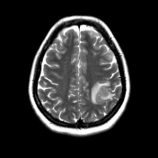
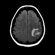
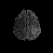
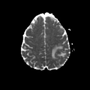
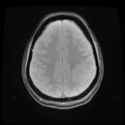

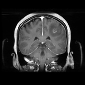
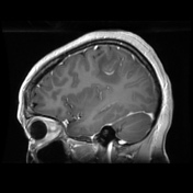
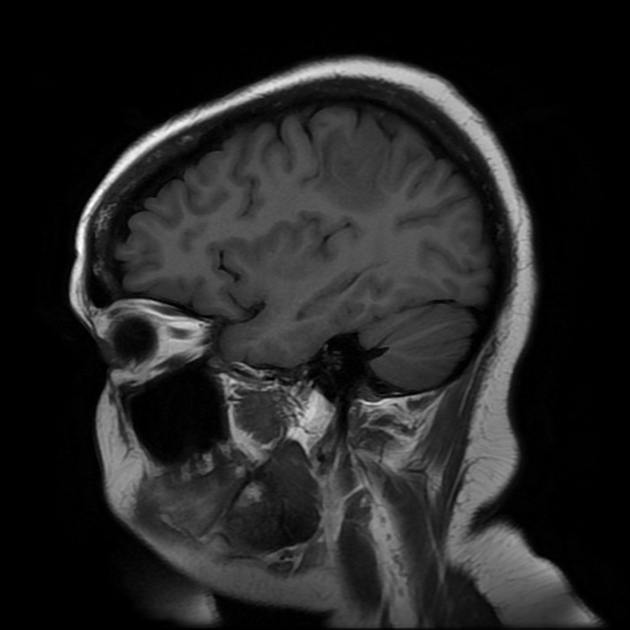
Edematous gyri seen in the involved region with T2 and T2 FLAIR hyperintense signals confined tothe subcortical and deep white matter suggestive of vasogenic edema. Post contrast study reveals a gyriform pattern of enhancement. Isointense to subtle hyperintense signal is seen in diffusion weighted images with isointense signals in ADC map.
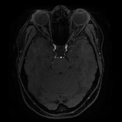
The angiogram of the circle of Willis was unremarkable apart from focal vasodilation in the afflicted site likely secondary to reperfusion changes.
In a known patient of Takayasu arteritis and the nature of acute onset symptoms with no other ancillary clinical features like fever, possibility of a subacute infarction was considered.

Reviewing the initial imaging findings at the time of first presentation.
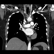
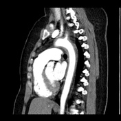
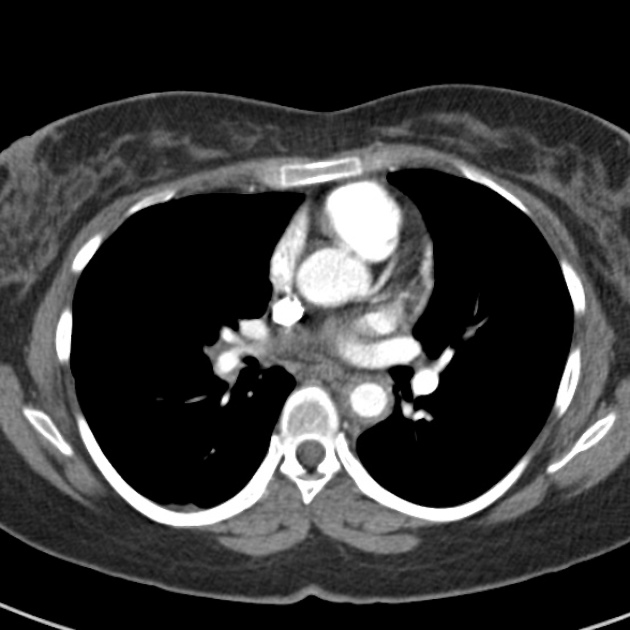
Circumferential mural thickening is seen involving the arch and descending thoracic aorta, extending to involve the origins of all the great vessels with mild areas of stenosis in proximal segments of the subclavian arteries.
Case Discussion
Classically Takayasu arteritis involves large vessels, which include the largest artery in the body the aorta, and the major vessels originating from it. The association of cerebral findings is uncommon in large vessel vasculitis but has been reported.


 Unable to process the form. Check for errors and try again.